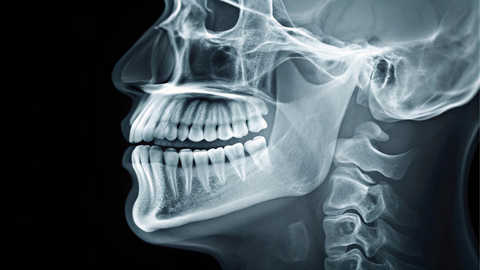
What Is a Cephalometric (Ceph) X-Ray?
A cephalometric X-ray is a standardized, side-view (lateral) dental radiograph of the head and face. It captures the skull, jaw bones, teeth, facial profile, and airway in a single image. Ceph X-rays are most commonly used in orthodontics because they allow for precise evaluation of how the teeth, jaws, and facial structures relate to one another.
Why We Use Cephalometric X-Rays
Cephalometric X-rays are used to evaluate the relationship between the jaws, teeth, and skull, as well as to assess facial growth and development. They help diagnose bite and jaw alignment issues and are especially valuable for monitoring changes over time by comparing images taken at different stages of treatment. This makes them an essential tool for planning and tracking orthodontic care.
Why Cephalometric X-Rays Are Important
Ceph X-rays provide the precise measurements needed for accurate orthodontic treatment planning. They help predict future growth patterns, particularly in younger patients, and assist in determining whether treatment may involve braces or aligners, jaw growth modification, or surgical intervention. These images also create a reliable baseline record, allowing progress to be measured clearly throughout treatment.
What Can Be Seen on a Cephalometric X-Ray
A cephalometric X-ray offers detailed insight into facial and dental alignment, including:
- Position and angulation of teeth
- Upper and lower jaw relationships (maxilla and mandible)
- Facial skeletal structure and overall profile
- Bite discrepancies such as overbite, underbite, and open bite
- Growth patterns of the face and jaws
- Airway space assessment
- Soft tissue profile, including lips, chin, and nose outline
At Bridgeport Smiles, cephalometric X-rays help us plan orthodontic treatment with clarity and confidence ensuring efficient care tailored to your unique facial structure and growth.